Beranda
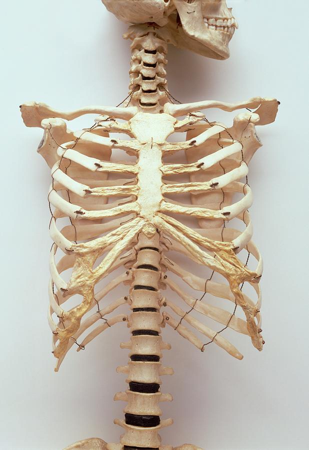
/ Anatomy Diagram Rib Area - 5vgd4k2coedz3m - The head only articulates with the body of the t1 vertebra and therefore only one articulatory surface is present.
Anatomy Diagram Rib Area - 5vgd4k2coedz3m - The head only articulates with the body of the t1 vertebra and therefore only one articulatory surface is present.
Insurance Gas/Electricity Loans Mortgage Attorney Lawyer Donate Conference Call Degree Credit Treatment Software Classes Recovery Trading Rehab Hosting Transfer Cord Blood Claim compensation mesothelioma mesothelioma attorney Houston car accident lawyer moreno valley can you sue a doctor for wrong diagnosis doctorate in security top online doctoral programs in business educational leadership doctoral programs online car accident doctor atlanta car accident doctor atlanta accident attorney rancho Cucamonga truck accident attorney san Antonio ONLINE BUSINESS DEGREE PROGRAMS ACCREDITED online accredited psychology degree masters degree in human resources online public administration masters degree online bitcoin merchant account bitcoin merchant services compare car insurance auto insurance troy mi seo explanation digital marketing degree floridaseo company fitness showrooms stamfordct how to work more efficiently seowordpress tips meaning of seo what is an seo what does an seo do what seo stands for best seotips google seo advice seo steps, The secure cloud-based platform for smart service delivery. Safelink is used by legal, professional and financial services to protect sensitive information, accelerate business processes and increase productivity. Use Safelink to collaborate securely with clients, colleagues and external parties. Safelink has a menu of workspace types with advanced features for dispute resolution, running deals and customised client portal creation. All data is encrypted (at rest and in transit and you retain your own encryption keys. Our titan security framework ensures your data is secure and you even have the option to choose your own data location from Channel Islands, London (UK), Dublin (EU), Australia.
Anatomy Diagram Rib Area - 5vgd4k2coedz3m - The head only articulates with the body of the t1 vertebra and therefore only one articulatory surface is present.. In this image you can see very clearly that the flank area is just above the hips and groin area. We are pleased to provide you with the picture named heart, lung, diaphragm and ribs location.we hope this picture heart, lung, diaphragm and ribs location can help you study and research. Each pair is numbered based on their attachment to the sternum, a bony process at the front of the rib cage which serves as an anchor point. The head only articulates with the body of the t1 vertebra and therefore only one articulatory surface is present. The rib cage is collectively made up of long, curved individual.
The superior surface is unique in that it is marked by two grooves that allow. See more ideas about anatomy, anatomy study, rib cage anatomy. Rib cage, in vertebrate anatomy, basketlike skeletal structure that forms the chest, or thorax, and is made up of the ribs and their corresponding attachments to the sternum (breastbone) and the vertebral column.the rib cage surrounds the lungs and the heart, serving as an important means of bony protection for these vital organs.in total, the rib cage consists of the 12 thoracic vertebrae and. Rib cage pain may be sharp, dull, or achy pain felt at or below your chest or above your navel on either side. They move up and down in the anterior chest, allowing for full chest expansion.
Human Rib Cage Photograph By Dorling Kindersley Uig from images.fineartamerica.com It also covers some common conditions and injuries that can affect the back. Don't be fooled their long, curved shape! The costal cartilages of the second through tenth ribs connect to the body of the sternum to form the bulk of the rib cage. True ribs, false and floating. These are short ribs that do not attach to. The liver weighs about 1,500 grams (~3 pounds) in average 1, 14. Rib cage, in vertebrate anatomy, basketlike skeletal structure that forms the chest, or thorax, and is made up of the ribs and their corresponding attachments to the sternum (breastbone) and the vertebral column.the rib cage surrounds the lungs and the heart, serving as an important means of bony protection for these vital organs.in total, the rib cage consists of the 12 thoracic vertebrae and. The rib cage is a bony structure found in the chest (thoracic cavity).
The liver weighs about 1,500 grams (~3 pounds) in average 1, 14.
For more anatomy content please follow us and visit our website: It is made up of 12 pairs of ribs. Sharp lines along the lower margin of the ribs, rib overlying shadows) may mimic pleural and extrapleural disease on frontal chest radiographs. See more ideas about anatomy, anatomy study, rib cage anatomy. Redness and warmth to the sternal and rib area may also be noted. Rib 1 is also flattened horizontally. The anatomy of the human ribs is made up of 24 ribs which are parted in 12 pairs (each on the left and right side of the chest wall), with the sternum, metasternum (the xiphoid process), and the costal cartilages all situated at the anterior of the chest wall, followed by the thoracic vertebrae on the posterior of the chest wall. True ribs, false and floating. The 11 th and 12 th ribs, known as floating ribs, are not attached in any way to the sternum; The rib cage is collectively made up of long, curved individual. Its functions are to protect the thoracic organs from trauma and also form the bony attachment for various muscles. It also covers some common conditions and injuries that can affect the back. Rib bones are not classified as long bones.instead, anatomists classify the ribs as flat bones, and they are located within the axial skeleton.together with the sternum, thoracic vertebrae, and costal cartilages, the ribs form the thoracic cage, also called the bony thorax.
It only slightly enters where the ribs are located. The liver weighs about 1,500 grams (~3 pounds) in average 1, 14. Don't be fooled their long, curved shape! Rib 2 is thinner and longer than rib 1, and has two articular facets on the head as normal. In this image you can see very clearly that the flank area is just above the hips and groin area.
Premium Vector Human Skeleton Structure Skull Spine Rib Cage Pelvis Joints Anatomy And Medicine 3d Icon Set from image.freepik.com It is also the center around which the superior 10 ribs directly or indirectly attached. For more anatomy content please follow us and visit our website: Each pair is numbered based on their attachment to the sternum, a bony process at the front of the rib cage which serves as an anchor point. They move up and down in the anterior chest, allowing for full chest expansion. The head only articulates with the body of the t1 vertebra and therefore only one articulatory surface is present. Rib bones are not classified as long bones.instead, anatomists classify the ribs as flat bones, and they are located within the axial skeleton.together with the sternum, thoracic vertebrae, and costal cartilages, the ribs form the thoracic cage, also called the bony thorax. The anatomy of the human ribs is made up of 24 ribs which are parted in 12 pairs (each on the left and right side of the chest wall), with the sternum, metasternum (the xiphoid process), and the costal cartilages all situated at the anterior of the chest wall, followed by the thoracic vertebrae on the posterior of the chest wall. It only slightly enters where the ribs are located.
True ribs, false and floating.
Labeled human stomach anatomy 6 photos of the labeled human stomach anatomy labeled human stomach anatomy, labeled human stomach anatomy picture, labeled human stomach diagram, stomach model labeled, stomach model labeled picture, stomach, labeled human stomach anatomy, labeled human stomach anatomy picture. Thus, the cartilage of rib 10 attaches to the cartilage of rib 9, rib 9 then attaches to rib 8, and rib 8 is attached to rib 7. The bones of the rib cage are the sternum, the 12 thoracic vertebrae and the 12 pairs of ribs. The anatomy of the human ribs is made up of 24 ribs which are parted in 12 pairs (each on the left and right side of the chest wall), with the sternum, metasternum (the xiphoid process), and the costal cartilages all situated at the anterior of the chest wall, followed by the thoracic vertebrae on the posterior of the chest wall. Rib 1 is also flattened horizontally. The ribs partially enclose and protect the chest cavity, where many vital organs (including the heart and the lungs) are located. On the trunk of the body in the thoracic area, the shoulder in general is the acromial, while the curve of the shoulder is the deltoid. The rib cage is a bony structure found in the chest (thoracic cavity). Anatomy of the rib cage diagram anatomy of the rib cage diagram in this image, you will find thoracic vertebrum, costochondral joint, costal cartilage, costal margin, costal arch, thoracic vertebrum, xiphoid process, xiphisternal joint, body, manubrial sternal joint, manubrium, the sternal notch in it. Redness and warmth to the sternal and rib area may also be noted. Costochondritis can cause sharp, stabbing rib pain and tenderness to any of the first three ribs. Just like in the manubrium, slight concave indentations in the lateral sides of the body of the sternum provide stronger attachment points for the costal cartilages to prevent rib separation. Rib cage pain may be sharp, dull, or achy pain felt at or below your chest or above your navel on either side.
It is also the center around which the superior 10 ribs directly or indirectly attached. The top edge of the manubrium has a depression called the suprasternal or jugular notch. Each pair is numbered based on their attachment to the sternum, a bony process at the front of the rib cage which serves as an anchor point. The primary responsibilities of the ribcage involve protecting the thoracic visceral organs, enclosing the thoracic visceral organs, and is included. The following diagram shows the anterior chest again, with the lobes of the lungs included.
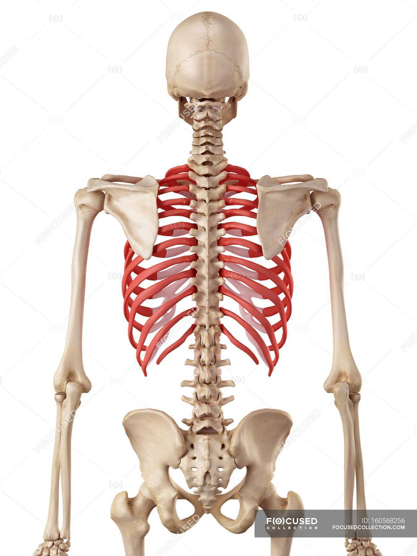
Human Rib Cage Anatomy Human Physiology Osteology Stock Photo 160568256 from st.focusedcollection.com The rib cage is a bony structure found in the chest (thoracic cavity). The 11 th and 12 th ribs, known as floating ribs, are not attached in any way to the sternum; Rib bones are not classified as long bones.instead, anatomists classify the ribs as flat bones, and they are located within the axial skeleton.together with the sternum, thoracic vertebrae, and costal cartilages, the ribs form the thoracic cage, also called the bony thorax. Rib 2 is thinner and longer than rib 1, and has two articular facets on the head as normal. Rib 1 is also flattened horizontally. The ribs partially enclose and protect the chest cavity, where many vital organs (including the heart and the lungs) are located. Don't be fooled their long, curved shape! The head only articulates with the body of the t1 vertebra and therefore only one articulatory surface is present.
These are short ribs that do not attach to.
The costal cartilages of the second through tenth ribs connect to the body of the sternum to form the bulk of the rib cage. Thus, the cartilage of rib 10 attaches to the cartilage of rib 9, rib 9 then attaches to rib 8, and rib 8 is attached to rib 7. The rib cage is a bony structure found in the chest (thoracic cavity). Abdominal anatomy muscles 12 photos of the abdominal anatomy muscles abdominal muscles anatomy and function, abdominal muscles anatomy diagram, abdominal muscles cross sectional anatomy, deep abdominal muscles anatomy, lateral abdominal muscles anatomy, human anatomy, abdominal muscles anatomy and function, abdominal. Redness and warmth to the sternal and rib area may also be noted. Rib cage pain may be sharp, dull, or achy pain felt at or below your chest or above your navel on either side. It is made up of 12 pairs of ribs. Anatomy of the rib cage diagram anatomy of the rib cage diagram in this image, you will find thoracic vertebrum, costochondral joint, costal cartilage, costal margin, costal arch, thoracic vertebrum, xiphoid process, xiphisternal joint, body, manubrial sternal joint, manubrium, the sternal notch in it. Rib 2 is thinner and longer than rib 1, and has two articular facets on the head as normal. The 11 th and 12 th ribs, known as floating ribs, are not attached in any way to the sternum; Ribs 11 and 12 do not have necks or tubercles and the anterior tips of their bodies lack an articular surface. The ribs partially enclose and protect the chest cavity, where many vital organs (including the heart and the lungs) are located. The seven superior pairs of ribs connect directly to the sternum via the costal cartilage and are collectively known as the true ribs.